Welcome to Pink Door Imaging
Breast Imaging Center
Pink Door Imaging for Breast and Gynecological Imaging offers an integrated approach to women's diagnostic imaging delivering both gynecological and 3D breast imaging services in a caring, respectful boutique practice environment. Our goal is to educate, relieve anxiety and guide our referring physicians and patients in making the best decision in clinical management.

MEDICAL DIRECTOR AND OWNER
MAHESH K. SHETTY MD
Pink Door Imaging P-LLC was founded by Dr. Mahesh Shetty. He is a fellow of the American College of Radiology, the Society of Breast Imaging and the American Institute of Ultrasound in Medicine. His expertise in breast and gynecological imaging has earned him distinction as a published thought leader, a clinical professor and the founder of a multinational nonprofit service organization, Woman’s Cancer Foundation, which advocates for cost effective combined screening methodology for breast and Gynecological cancers in low resource countries.
- Specialized in Breast Cancer Screening and Gynecological Imaging
- Physician owned offering a personalized experience, opportunity to discuss findings in an interactive environment.
- Breast cancer screening and diagnosis performance metrics equals or exceeds established national performance benchmarks, low false positives with optimal cancer detection rate and positive biopsy rate.
- Easy access, location, appointment availability, screening mammograms done on a walk in and self-referral basis.
- Continuum of care, efficient and timely communication with patients and referring doctors for optimal patient care and follow up.
- Cost: Highly competitive. All major insurances are accepted. Negotiated rates are close to Medicare rates which translates to relatively low cost to patients who have not met their deductible. Cash prices are competitive.


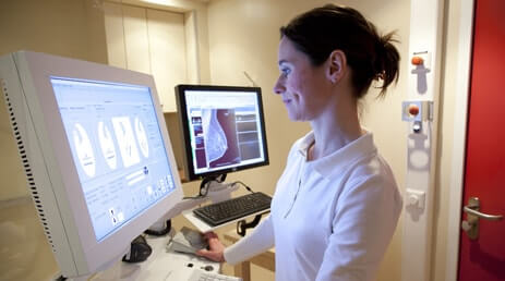
3D Mammography or Digital breast tomosynthesis
We have chosen the best 3D mammography system for your patients, one that supports a more reliable diagnosis and reduces the need for callbacks. Mammomat Inspiration with True Breast Tomosynthesis is the wide-angle option that helps us to be sure of diagnostic accuracy. Why go wide?
Because the width of the angle and the number of projections determine the quality of the resulting 3D image. Our wide-angle tomosynthesis acquires 25 images of the breast across a 50° angle, resulting in better depth resolution and tissue layer separation – and increased breast cancer detection rates.
Diagnostic Mammography
A diagnostic mammography is an x-ray exam that is performed when an abnormality is discovered during a mammogram. This exam is utilized to identify the size and location of the lesion. A diagnostic mammography may also help determine if the abnormality is benign or cancerous.
Dr. Shetty understands the stress patients face during this time and strives to offer appointments within 1-2 business days. All results will be thoroughly discussed with the patient following the exam. Woman’s Clinic uses the latest techniques and technology to perform all exams and procedures.
Breast Ultrasound
The breast ultrasound is a noninvasive exam that can be used to view lumps or any abnormalities that may not be seen in mammograms and other breast exams. There is no radiation involved in this exam and it is ideal for younger or pregnant patients. It is often used in place of a diagnostic mammography.
For patients with an elevated risk for breast cancer, the breast ultrasound exam can be used as a supplement to mammography. The ultrasound exam can easily find smaller lesions not typically found in clinical exams. The breast ultrasound can be combined with other exams to pinpoint and evaluate lesions.
Minimally Invasive Breast Biopsy
Dr. Shetty utilizes a state-of-the-art ultrasound or stereotactic guidance to perform a breast biopsy procedure. A minimally invasive biopsy is often the first approach to an abnormality in place of a surgical approach. The biopsy is performed at Woman’s Clinic as an outpatient procedure. Patients will be carefully placed under local anesthesia in preparation for the biopsy.
The procedure will help determine if the abnormality is cancerous. Dr. Shetty will educate the patient on all treatment options. The entire Woman’s Clinic staff is dedicated to creating a safe and comfortable environment for all patients.
Gynecological Ultrasound
A gynecological ultrasound, also known as a pelvic ultrasound, is utilized to diagnose symptoms in pelvic organs and structures. The exam uses high-frequency sound waves to create a clear image of the pelvic area. The ultrasound exam helps accurately diagnose and manage pelvis symptoms. Pelvic ultrasounds may be performed if pelvic abnormalities are found.
Dr. Shetty will identify the size and location of the abnormality. There is no radiation used in the gynecological ultrasound exam. The pelvic ultrasound can be used in place of an invasive surgical procedure. Dr. Shetty utilizes the most advanced equipment to perform all exams at Woman’s Clinic.
Second Opinions
Second opinions are provided. Dr. Shetty suggests patients drop off a CD of the images and reports to set up an appointment with our physicians to discuss their findings. Our physicians will provide recommended treatments and procedures. Dr. Shetty utilizes state-of-the-art technology to perform all exams at Woman’s Clinic.
Patients can find peace of mind with the help of our breast cancer specialists. It is important for patients to feel comfortable when embarking on their cancer journey. Dr. Shetty believes that patients should be fully educated in all diagnostic and treatment options.
3D Mammography or Digital breast tomosynthesis
Diagnostic Mammography
Breast Ultrasound
Minimally Invasive Breast Biopsy
Gynecological Ultrasound
Second Opinions
Breast Ultrasound

Breast Ultrasound is a non-radiological examination used as a supplemental modality to screen for breast cancer and to evaluate abnormalities that are seen on mammograms
Diagnostic Mammography

Diagnostic mammogram is a supervised mammographic examination that is tailored to evaluate specific breast symptoms and abnormal mammograms
Gynecological Ultrasound
Pelvic or gynecological ultrasound is a non-radiological modality used to evaluate the pelvic organs