Breast pain is a common problem that affects 70–80% of women at some point in their lives and is most frequent in premenopausal women. The most common underlying cause is fibrocystic change, a benign alteration of the terminal lobular ductal unit of the breast. Fibrocystic change encompasses all the changes associated with normal variations in breast parenchyma associated with changes in hormone levels.
To learn more about breast imaging in Houston, schedule a consultation with Dr. Shetty today.
Types of Breast Pain:
Breast pain can be cyclical in menstruating women, occurring before menstrual cycles making hormones a trigger. This type of pain tends to affect women in their 30’s and 40’s. These are often bilateral and worse in the upper outer portions of the breast. The breasts are tender and may feel lumpy. Cyclical pain is the most common type of breast pain. Treatment is symptomatic and there is no benefit in imaging. Non-cyclical breast pain is not related to menstrual cycles and may occur in one breast or in one quadrant of the breast.
Women in an older age or post-menopausal women generally report this type of pain. Breast pain is a common, often distressing problem among women. After significant disease is ruled out, most patients respond to simple reassurance. Others, however, require treatment because symptoms interfere with their lifestyle. Breast pain is generally not a symptom of breast cancer. Reports on the incidence of cancer in patients present with breast pain state the occurrence to be 0–3.2%. Treatment for most breast pain symptomatology can be reassurance, over-the-counter pain medications, or structural support.
As breast cancer awareness has increased, a concern that breast pain may indicate malignancy contributes to the trend of breast pain being the most common breast symptom causing a woman to consult her primary care physician or a breast surgeon. These studies report that after initial imaging, most women require no intervention after reassurance that their diagnostic imaging work-up is normal. Reports show the negative predictive value of mammography and ultrasound for patients with breast pain to be 100% in three studies.
When to Consult Your Physician
Seek a physician consultation to determine if you need additional testing, particularly when any of the following occur with breast pain:
• Breast pain is localized and not related to menstrual cycle
• When it is associated with a breast lump
• When it is associated with skin changes or itching
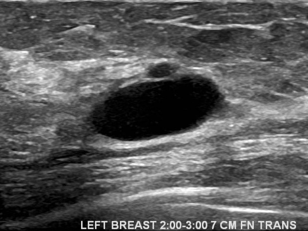
Your physician can order a breast ultrasound with or without a diagnostic mammogram. The choice depends on their assessment of the symptoms and any additional findings. These imaging studies will help to exclude an underlying breast problem. A mammogram may show a mass at the site of breast pain in women with a breast lump, while an ultrasound will be able to determine if the mass is a fluid filled cyst or a solid tumor. Fluid filled cysts are left alone unless they are painful in which case these can be drained with or without ultrasound guidance as an office based procedure. (Figure 1). If the examining doctor identifies a solid tumor, the performance of an outpatient biopsy where a ultrasound directs a needle to the tumor can occur. This procedure depends on the characteristics of the breast tumor.
A rare specific cause of breast pain that is relates to a palpable cord like lump is Mondor’s disease. In this condition there is blockage of a vein by a clot leading to presence of painful cord like lump. (Figure 2). This is a self-limiting condition that responds to anti-inflammatory analgesics. An ultrasound and a mammogram can readily diagnose this condition. (Shetty MK, Watson AB, https://www.ncbi.nlm.nih.gov/pubmed/11566698)
How to Manage Breast Pain ?
Breast pain is usually self-limited and is not typically a symptom of malignant pathologic disease. Treatment for most breast pain symptoms can be reassurance, over-the-counter pain medications, or structural support. The most important factors in the evaluation and treatment of breast pain consist of a thorough history, physical, and radiologic evaluation.
These evaluations can reassure the patient that she does not have breast cancer. In the 15% of mastalgia patients who have life-altering pain and still request treatment, therapy may consist of a well-fitting bra, a decrease in dietary fat intake, and discontinuance of oral contraceptives or hormone replacement therapy. Those women still resistant to therapy may experience relief from evening primrose oil supplements, bromocriptine, tamoxifen, or GnRH analogues.
Predicting which treatment will be most useful for any woman may be challenging. When severe and intolerable cyclical mastalgia shows to benefit from Vitamin E (1200 IU daily) and evening primrose oil (3000 mg daily) (1). Severe cases of mastalgia or breast pain may respond to Danazol treatment which is a prescription medication (3). Bromocriptine, also a prescription medication has been studied in treatment of cyclical breast pain with some benefit (4).
Breast Imaging in Houston
At Pink Door Imaging we pride ourselves in delivering a comfortable, state-of-the-art experience for our patients. For more information on breast imaging in Houston, don’t hesitate to get in touch with Dr. Shetty today.
Resources:
1. https://radiopaedia.org/articles/fibrocystic-change-breast
2. https://www.ncbi.nlm.nih.gov/pubmed/20359269
3. https://www.ncbi.nlm.nih.gov/pubmed/1548647
4. http://journals.sagepub.com/doi/abs/10.1177/003693308002500449?journalCode=scma
5. https://www.ncbi.nlm.nih.gov/pubmed/7816407
6. https://www.tandfonline.com/doi/pdf/10.3810/pgm.1997.11.369?needAccess=true
7. Mastalgia. Academic Radiology, 2017-03-01, Volume 24, Issue 3, Pages 345-349
Figure 1 A. Mammogram in a patient with breast pain and a lump shows a mass at the site.
Figure 1B. Ultrasound in the same patient demonstrates a fluid filled cyst.
Figure 2. A patient presenting with breast pain and a palpable cord like breast lump. Ultrasound shows a distended vein characteristic of Mondor’s disease of the breast.


