
A pelvic ultrasound can be an essential component of an individual’s overall health. This procedure has the ability to help a medical health professional diagnose and identify many potential health issues having to do with reproductive and general health – including breast cancer screening. Below we look at: is a pelvic ultrasound painful?
One of the great things about this procedure is that, for most people, it’s entirely painless. This means that a great majority of individuals will be able to undergo this procedure in an entirely harmless manner.
Is a Pelvic Ultrasound Painful?
If you’re interested in having a pelvic ultrasound performed or already have one scheduled, take a look at this guide to the procedure.
Pelvic Ultrasound Basics
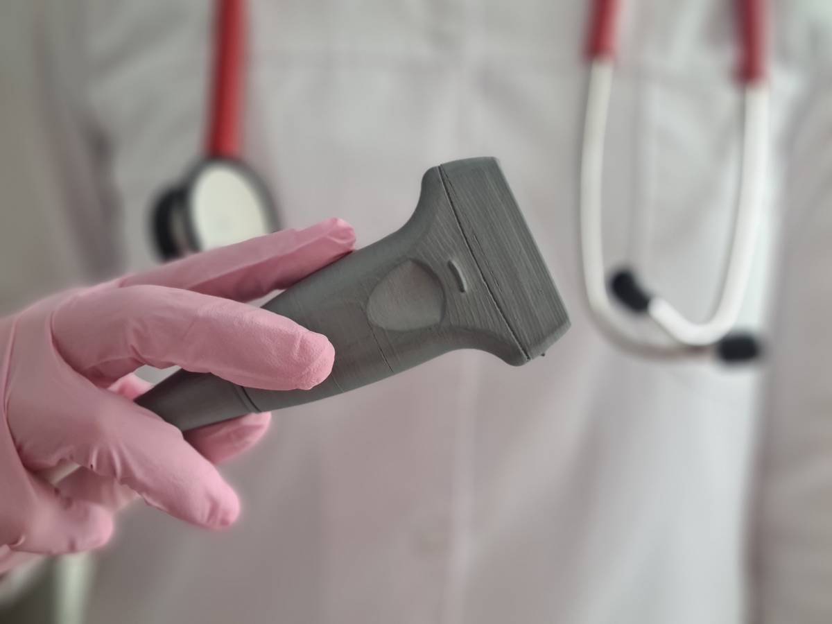
A pelvic ultrasound is a specific type of ultrasound intended to diagnose diseases and conditions affecting the health of the uterus and other reproductive organs. During this type of ultrasound, the ultrasound probe is placed in the vagina of the patient.
There are several different types of conditions and abnormalities that a pelvic ultrasound can help to detect. Some of these include:
- Cancers of the uterus, ovaries, and other parts of the reproductive system
- Certain types of infections, such as pelvic inflammatory disease
- Endometriosis
- Certain non-cancerous growths that may be affecting the ovaries (examples include cysts and fibroids).
- Twisted ovaries
- Pregnancy outside of the uterus
- Birth defects
Pelvic ultrasounds can be highly useful at identifying conditions such as these either prior or during their full maturation. From diagnosis, a gynecologist can help to develop a plan for further treatment.
How the Test Transpires
During a pelvic ultrasound, a patient lies down on a table with their knees bent. Your feet may be contained in stirrups. After the correct position is assumed, your physician will introduce a probe into the vagina. In most cases, this can produce some mild discomfort but very rarely pain.
The probe is covered with a condom and some gel. At this point, the true ultrasound process begins, which entails the following:
- The probe transmits sound waves and records the reflections of the waves of the structures of this area of the body. This, in turn, creates an image of the body parts.
- The image that is created through the ultrasound process is displayed on the ultrasound machine. Both doctor and patient will have a full view of the image created on the ultrasound machine in most cases.
- The provider will be able to gently move the probe around the area to further investigate the pelvic organs of the patient.
In some instances, the physician will need to apply a special substance in order to clarify the image results of the ultrasound. This substance is called a saline infusion sonography, or SIS.
A Pain-free Procedure
Preparing for an ultrasound is a fairly rudimentary process and simply involves stripping from the waist down and wearing a hospital gown on the upper body. In addition, your physician will usually request that you fully empty your bladder in order to facilitate the testing process.
A pelvic ultrasound is a primarily pain-free procedure. The only times that pain should be experienced is if the patient has some kind of special condition that would cause pain, such as some kind of STI or other painful condition.
That being said, the pressure of the probe is liable to cause at least some small amount of discomfort for the patient due to the sensitive nature of the area of the body that is being covered. Preparing for this discomfort mentally can give you.
Your Results
In the case of normal results, no further action is required on the part of the patient or the physician. In the case of abnormal results, your gynecologist will understand the necessary next steps for the patient to take charge of their medical situation.
The nature of these steps will depend heavily on the nature of the abnormality that is exposed during the process. As these abnormalities can vary heavily in terms of severity, it is somewhat hard to predict what the exact nature of the patient’s further treatment will be.
Ultrasound results can come in handy in other areas of a patient’s medical life as well; holding on to yours may save you time in further healthcare scenarios in the future.
The Ultrasound Experts of Houston
Pink Door Imaging and its passionate team are completely dedicated to the safety and health of our clients. If you’re interested in having a pelvic ultrasound performed or would like to discuss the details of how this procedure works, contact our office in order to schedule a consultation.