Abnormal uterine bleeding is a common gynecological complaint for which women seek consultation with a Gynecologist. Following an examination, a pelvic ultrasound is routinely performed. The main purpose of the ultrasound examination is to determine a cause for the abnormal bleeding. Based on the findings, the gynecologist will plan an optimal treatment.
Abnormal Uterine Bleeding: Bleeding from the uterus that differs in frequency, regularity, duration, or amount from normal uterine bleeding in the absence of pregnancy.
When is bleeding abnormal?
Bleeding in any of the following situations is considered abnormal uterine bleeding:
• Bleeding or spotting between periods
• Bleeding or spotting after sex
• Heavy bleeding during your period
• Menstrual cycles that are longer than 38 days or shorter than 24 days
• “Irregular” periods in which cycle length varies by more than 7–9 days
• Bleeding after menopause
Some of the causes of abnormal bleeding include the following:
• Problems with ovulation
• Fibroids and polyps
• A condition in which the endometrium grows into the wall of the uterus
• Bleeding disorders
• Problems linked to some birth control methods, such as an intrauterine device (IUD) or birth control pills
• Miscarriage
• Ectopic pregnancy
• Certain types of cancer, such as cancer of the uterus
Investigation of abnormal uterine bleeding
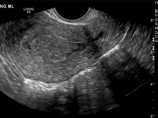
A pelvic ultrasound is always performed as the first line of testing in women who present with a complaint of abnormal uterine bleeding. Ultrasound very clearly visualizes the endometrial lining (Figure 1) and identifies potential causes for the bleeding. The causes of the abnormal bleeding that is diagnosed with pelvic ultrasound include:
Abnormalities of the endometrium (inner lining of the uterus)
The endometrial cavity can contain a tumor that is readily identified by an ultrasound exam. These may include endometrial polyps that are non-cancerous growths of the uterine lining. These tumors are readily diagnosed on pelvic ultrasound (Figure 2). Pelvic ultrasound with Doppler will reveal a stalk with blood supply to the polyp, Treatment is often through a simple procedure called hysteroscopy where a scope is introduced into the uterine cavity and the polyp is removed under direct visualization. A fibroid can grow inside the endometrial lining and cause abnormal bleeding. Distinction between a polyp and a fibroid is often possible based on the ultrasound features but is not critical in the management since both conditions are similarly treated.
Abnormalities of the myometrium (Uterine muscle)
Uterine fibroids are common benign non-cancerous tumors of the uterine muscle. These are seen in the uterine muscle and occur in more than a third of women during menstrual age. The size can vary from that of a dime to a grapefruit. (Figure 2 A and B). There is frequently more than one fibroid. If not symptomatic, they are left alone. The fibroids that are impinging up on the endometrial lining can cause abnormal bleeding. Such fibroids are called submucosal fibroids. A radiologist with expertise in Pelvic ultrasound can describe in detail the information that a gynecologist needs to plan optimal mode of intervention. A pelvic ultrasound accurately identifies and diagnoses fibroids and can determine if it is projecting into the endometrial lining. All fibroids are non-cancerous, about 1 in 1000 may be the cancerous counterpart called leiomyosarcoma. Ultrasound and other imaging tests are not able to precisely characterize the cancerous counterpart of a fibroid. There are multiple treatment options for women in whom fibroids cause symptoms and the gynecologist will discuss surgical and non-surgical options with the patient. The latter is important in those who seek to preserve the uterus.
Adenomyosis is a condition where the endometrial lining of the uterus grows in to the uterine muscle. This leads to enlargement of the uterus and focal masses in the myometrium. Adenomyosis can cause abnormal uterine bleeding and is often accompanied by painful menstruation and or painful intercourse. A combination of painful and increased bleeding during a menstrual cycle may be caused by this condition that has been reported in up to a third of all women. A pelvic ultrasound identifies focal and diffuse adenomyosis. Localized adenomyosis can be mistaken for a fibroid. The distinction is important since focal adenomyosis unlike fibroids are not amenable to surgical removal. It is important to seek radiologists specialized in Gynecological Imaging so as to have an accurate diagnosis, so management can then be tailored based on the cause of the abnormal bleeding.
Malposition of an IUD
This can cause abnormal bleeding with or without associated pain. An endovaginal ultrasound with 3D imaging accurately localizes the IUD and ensures that it is in satisfactory position within the endometrial cavity (Figure 3). If a portion or all the IUD is embedded in the uterine muscle, patient may experience symptoms and the IUD may not be effective. This is readily diagnosed on a pelvic ultrasound exam and may necessitate removal and replacement.
Abnormal bleeding in a Post-menopausal woman
Abnormal bleeding in a post-menopausal woman must be promptly investigated with a pelvic ultrasound. The endometrial lining is readily assessed. The radiologist inspects the endometrial lining and measures the thickness of the endometrium. The measurement of the endometrial lining will decide the need for an endometrial biopsy. About 10% of women with endometrial bleeding will have endometrial cancer (Figure 4). Ultrasound helps to triage a patient into a group that needs biopsy and a group that does not require it. There are other abnormalities of the endometrium that are diagnosed with an endovaginal pelvic ultrasound. These include changes suggestive of cystic hyperplasia or a growth in the endometrium called a polyp. Endometrial cancer is suspected when the endometrium is markedly thickened and or irregular and when the junction of the endometrium and myometrium is indistinct. Post-menopausal women with abnormal bleeding should promptly seek attention to identify an underlying cause, a pelvic ultrasound is an integral part of such an assessment. Endometrial cancer is the underlying cause in a small but significant number of patients and has excellent prognosis if diagnosed at an early stage when the cancer is still confined to the endometrial lining.
Resources:
https://www.acog.org/Patients/FAQs/Abnormal-Uterine-Bleeding
https://www.womenshealth.gov/a-z-topics/uterine-fibroids
https://www.nichd.nih.gov/health/topics/uterine/conditioninfo/default
https://rarediseases.info.nih.gov/diseases/8156/adenomyosis
Figure 1: Endovaginal Ultrasound shows the normal endometrial lining of the uterus
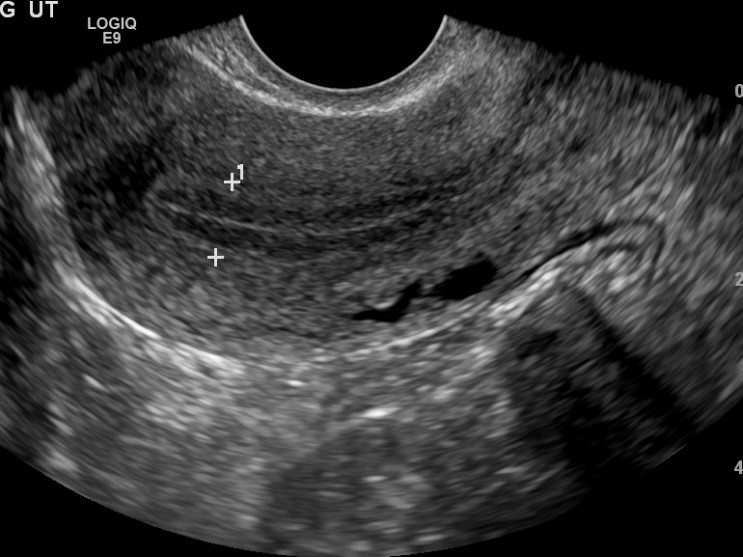
Figure 2 A. Pictorial representation of fibroids in the uterus. B. Endovaginal ultrasound shows a fibroid in the uterine muscle.
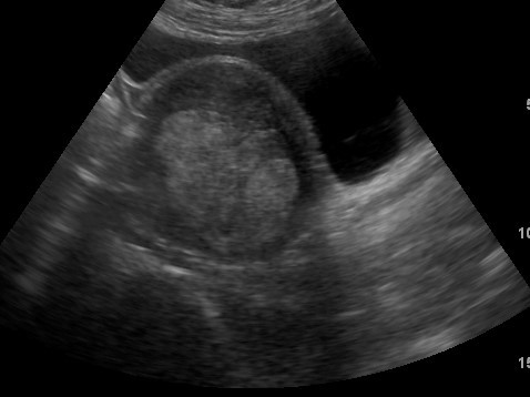
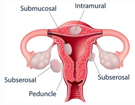
Figure 3. A 3D ultrasound shows that the IUD is in satisfactory location within the endometrial cavity
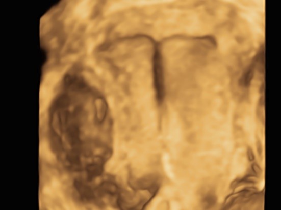
Figure 4: Endovaginal ultrasound shows a growth in the endometrial lining in a post-menopausal patient who presented with abnormal bleeding. At biopsy an endometrial cancer was found.