Pelvic pain in women, whether sudden in onset or of long standing is one of the most common symptoms that leads to a gynecologist visit. Following a physical examination and symptom assessment a pelvic ultrasound is often ordered. Ready availability, relatively low cost, and lack of ionizing radiation make ultrasonography an ideal imaging modality in women to assess the cause of the pelvic pain.
About the Exam
A Pelvic Ultrasound is typically performed by scanning over the lower abdomen, this is called as transabdominal pelvic ultrasound and requires a full bladder for optimal visualization of the uterus and ovaries. This is followed by a transvaginal ultrasound after the bladder is emptied. Transvaginal pelvic ultrasound provides a clear image of the ovaries, the uterus and the fallopian tubes. Typically, the examination takes less than thirty minutes. The radiologist reviews the images obtained by the ultrasound technologist. Experience and expertise of the radiologist is important in accurate diagnosis of the findings on a pelvic ultrasound exam.
Causes of Acute Pelvic Pain in Women and Diagnosis on a Pelvic Ultrasound Exam
In a woman of a reproductive age group early pregnancy is usually excluded by a urine/blood test. A pelvic ultrasound is the initial test ordered to exclude a gynecological cause for the pelvic pain. A pelvic ultrasound will readily identify an adnexal cause of the pain and help the gynecologist decide on the most appropriate treatment. Ultrasound findings will aid in deciding on medical conservative treatment and or the need for laparoscopic or surgical intervention.
The most common causes for an acute pelvic pain are:
Hemorrhagic cyst in the ovary: Cysts in the ovary are common and often physiologic, when small they do not cause any symptoms. When there is bleeding into a cyst they enlarge and cause pain. If the cyst leaks, there is spill of blood from the cyst into the pelvic cavity. This leads to pain of acute onset. A pelvic ultrasound particularly an endovaginal ultrasound identifies the cyst in the ovary, blood within it and in the pelvic cavity.
Pelvic infection also called Pelvic inflammatory disease or PID: Infection spreading to the fallopian tube can be a cause of acute onset of pain. This is caused by inflammation and distension of the fallopian tube. This may result in formation of an abscess in the pelvic cavity. Ultrasound is able to identify abnormal tubes and other signs of pelvic infection. Your gynecologist will decide on the management based on the findings on an ultrasound. An abscess will often need surgical intervention, earlier phases of infection can be managed medically. Ultrasound aids in the management by determining the state of the fallopian tubes and or presence of an abscess helping the gynecologist to manage such patients appropriately.
Endometriosis is a debilitating condition that can cause acute onset and chronic pain and painful menstrual cycles. In this condition the inner lining of the uterus called as the endometrium is found outside its normal location namely around the uterus and ovaries and in many other locations including the abdominal wall, urinary bladder. Endometrial tissue bleed during cycles and lead to formation of endometrial implants that can lead to infertility and bowel related problems. When they form blood filled cysts called as chocolate cysts, they are readily discovered on ultrasound. An endometrioma is a tumor like collection of blood clot in the pelvis. If large in a woman who is symptomatic surgical removal may be needed. Endometriomas are also sometimes hard to distinguish from ovarian neoplasms.
Ovarian torsion occurs when the blood supply to the ovary is compromised often due to a twist of the pedicle that brings in blood vessel to the ovary. A cyst or a tumor in the ovary is often the predisposing factor. A timely diagnosis is critical since this condition is a surgical emergency. Early surgical intervention based on accurate assessment by ultrasound can potential lead to salvaging of the affected ovary. A pelvic ultrasound will show the underlying tumor or cyst. The affected ovary is enlarged. Doppler techniques using ultrasound can assess blood flow to the ovary. In the initial phase venous blood flow is cut off followed by arterial blood flow. If not diagnosed early, oophorectomy or removal of the ovary may be the only option. It is therefore important to seek attention and obtain an ultrasound as quickly as possible. It is important to seek a center with expertise in Gynecological (Pelvic) ultrasound, so an accurate diagnosis may be made in a timely manner.
Tubal pregnancy can present with acute onset of pain caused by a growing pregnancy abnormally implanted in the fallopian tube. In these cases, a pregnancy test will be positive. An ultrasound will show no evidence of a pregnancy in the uterus. The fallopian tube will be found to be distended by the ectopic pregnancy and an associated blood clot. Ultrasound diagnoses ectopic pregnancy with a high degree of accuracy and can find signs of rupture. The gynecologist decides on the treatment based on the findings on a pelvic ultrasound. The size of the tubal pregnancy, symptoms experienced by the patient, evidence of a rupture or lack thereof on a pelvic ultrasound helps the gynecologist to decide on the optimal management of a patient with an ectopic pregnancy.
What if ultrasound is normal…a normal pelvic ultrasound in a woman with acute onset pain…. may need further testing which is usually a CT scan exam. Pelvic ultrasound is able to diagnose non-gynecological causes of pain such as appendicitis, diverticulitis or kidney stones. But these conditions are more accurately diagnosed on a CT scan.
Figure 1: Hemorrhagic cyst in the ovary in a patient with acute onset of pelvic pain
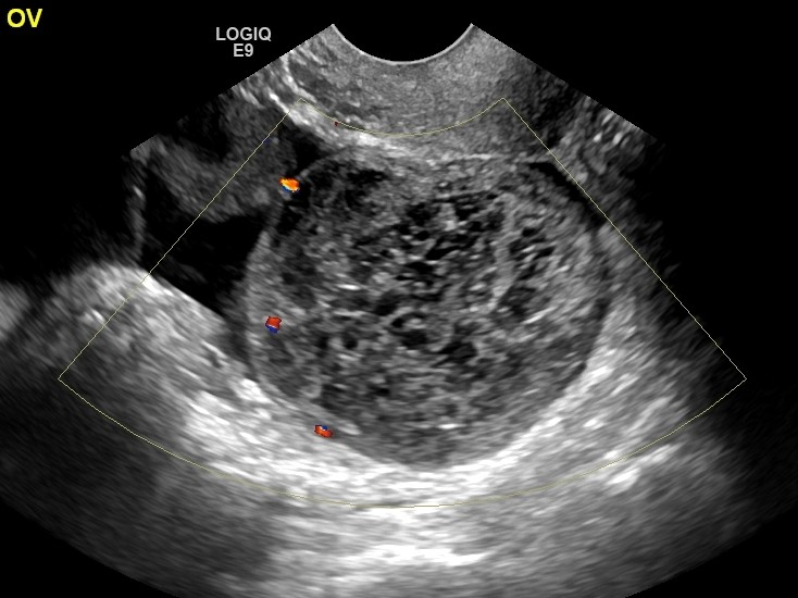
Figure 2. Ultrasound shows Pelvic inflammatory disease with a Tuboovarian abscess in a patient with acute onset of pelvic pain
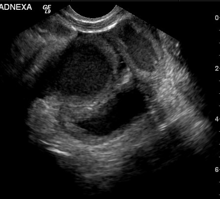
Figure 3. Ultrasound shows endometrioma in a patient with pelvic pain
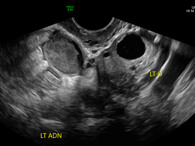
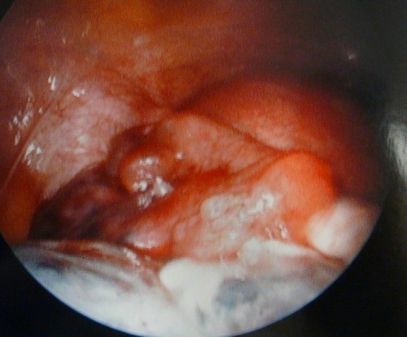
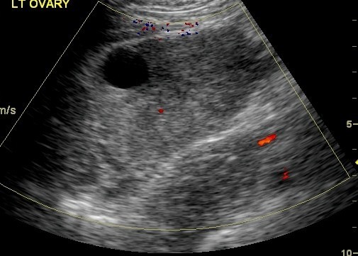
Figure 4. Ovarian torsion in a patient with acute onset of pelvic pain. Ultrasound and subsequent laparoscopy shows an ovary with no blood flow
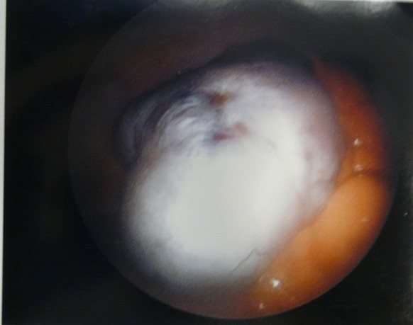
Restoration of blood flow seen after laparoscopic intervention indicating that the ovary has been salvaged.